Corsi e Congressi 2022-2023
MASTERCLASS DI ECOGRAFIA BEDSIDE - Bologna, 2-3/03/2023
MASTERCLASS DI ECOGRAFIA BEDSIDE - Bologna, 15-16/12/2022
MASTERCLASS DI ECOGRAFIA BEDSIDE - Bologna, 13-14/10/2022
MASTERCLASS DI ECOGRAFIA BEDSIDE (Addome superiore) - Bologna, 24/06/2022
Didattica e formazione (vedi curriculum)
RELAZIONI (2000-2017)
Di seguito sono riportate alcune tra le relazioni tenutesi negli ultimi anni.
LEZIONI E INSEGNAMENTI (2005-2013)
Semeiotica Internistica in Gastroenterologia nell'ambito della Scuola di Specializzazione in Gastroenterologia presso la sede di Roma dell’Università Cattolica del Sacro Cuore della Facoltà di Medicina e Chirurgia "A. Gemelli" (dal 2013)
Master Universitario di II Livello in Ecografia Internistica dell’Università degli Studi La Sapienza di Roma
Oncologia Medica (III Anno), corso integrato di tecniche infermieristiche applicate alla medicina clinica specialistica, Corso di Laurea in Infermieristica, Università degli Studi di Bologna (docenza fino al 2009)
Medicina Interna (II Anno), Scuola di Specializzazione in Gastroenterologia, Università Cattolica del Sacro Cuore di Roma (docenza fino al 2009)
Semeiotica clinica e strumentale in Medicina Interna (II Anno), integrativo di: Medicina Interna - Semeiotica medica Strumentale, Scuola di Specializzazione in Medicina dello Sport, Università degli Studi di Bologna (docenza fino al 2008)
Gastroenterologia (Fisiopatologia) presso la Scuola di Specializzazione in Gastroenterologia dell'Università Cattolica di Roma (docenza fino al 2005)
PUBBLICAZIONI
Le pubblicazioni di maggior rilevanza scientifica sono riportate su PubMed e visualizzabili all’indirizzo: http://www.ncbi.nlm.nih.gov/sites/entrez?cmd=search&db=pubmed&term=Arienti%20V%5Bau%5D&dispmax=50
PUBBLICAZIONI
RECENTI
-----------------------------------------------------------------------------------------------------------------------------------------------------
-----------------------------------------------------------------------------------------------------------------------------------------------------
Dig Liver Dis. 2016 Feb.
Percutaneous real-time sonoelastography as a non-invasive tool for the characterization of solid focal liver lesions: A prospective study.
Cesario V1, Accogli E2, Domanico A2, Di Lascio FM3, Napoleone L4, Gasbarrini A5, Arienti V2.
Abstract
BACKGROUND:
Real-time sonoelastography is currently used for the characterization of superficial solid lesions such as thyroid and breast masses. This study evaluates the usefulness of percutaneous sonoelastography for the characterization of solid focal liver lesions.
METHODS:
30 out of 43 patients with 38 known liver lesions were included in a prospective, diagnostic study. Qualitative analysis (pattern of deformation, elasticity type of liver tumour) and semi-quantitative measurements (strain ratio, hardness percentage, histogram) were evaluated. Sensitivity, specificity, positive and negative predictive values were calculated and the area under the receiver operating characteristics curve was constructed.
RESULTS:
Patterns A and C-D are specific of benign lesions and metastases respectively. The patterns for haemangiomas, focal nodular hyperplasia and metastases were significantly different to each other in terms of strain ratio, hardness percentage and histogram (p<0.05). A statistically significant difference (p<0.001) was observed between the median values of the 3 measured parameters for benign (1.02; 12%; 47) and malignant lesions (1.66; 65%; 20.5) respectively. The area under the receiver operating characteristics curve values for strain ratio, hardness percentage and histogram were 0.88, 0.89, and 0.86 respectively for cut-off values of 1.2, 45, and 30.
CONCLUSIONS:
By percutaneous sonoelastography it is possible to differentiate benign versus malignant focal liver lesions, metastases in particular, with good diagnostic performance.
Copyright © 2015 Editrice Gastroenterologica Italiana S.r.l. Published by Elsevier Ltd. All rights reserved.
KEYWORDS:
Benign and malignant focal liver lesions; Elastography; Hardness
-----------------------------------------------------------------------------------------------------------------------------------------------------
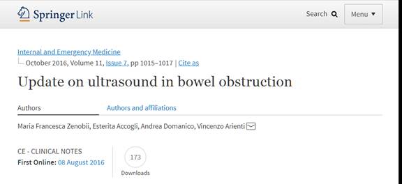
Intern Emerg Med. 2016 Mar
Update on bedside ultrasound (US) diagnosis of acute cholecystitis (AC).
Zenobii MF1, Accogli E1, Domanico A1, Arienti V2.
Abstract
Acute cholecystitis (AC) represents a principal cause of morbidity worldwide and is one of the most frequent reasons for hospitalization due to gastroenteric tract diseases. AC should be suspected in presence of clinical signs and of gallstones on an imaging study. Upper abdominal US represents the first diagnostic imaging step in the case of suspected AC. Computed tomography (CT) with intravenous contrast (IV) or magnetic resonance imaging (MRI) with gadolinium contrast and technetium hepatobiliary iminodiacetic acid (Tc-HIDA) can be employed to exclude complications. US examination should be performed with right subcostal oblique, with longitudinal and intercostal scans. Normal gallbladder US findings and AC major and minor US signs are described. Polyps, sludge and gallbladder wall thickening represent the more frequent pitfalls and they must be differentiated from stones, duodenal artifacts and many other non-inflammatory conditions that cause wall thickening, respectively. By means of bedside ultrasound, the finding of gallstones in combination with acute pain, when the clinician presses the gallbladder with the US probe (the sonographic Murphy's sign), has a 92.2 % positive predictive value for AC. In our preliminary experience, bedside US-performed by echoscopy (ES) and/or point-of-care US (POCUS) demonstrated good reliability in detecting signs of AC, and was always integrated with physical examination and performed by a skilled operator.
KEYWORDS:
Acute cholecystitis (AC); Bedside ultrasound (US); Diagnosis
-----------------------------------------------------------------------------------------------------------------------------------------------------
Intern Emerg Med. 2014 Oct
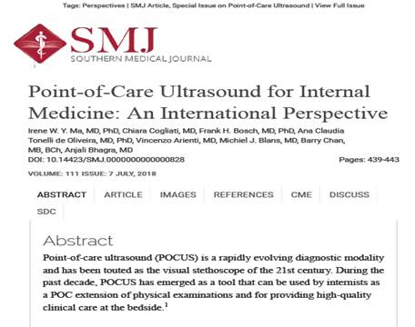
Bedside ultrasonography (US), Echoscopy and US point of care as a new kind of stethoscope for Internal Medicine Departments: the training program of the Italian Internal Medicine Society (SIMI).
Arienti V1, Di Giulio R, Cogliati C, Accogli E, Aluigi L, Corazza GR; Ultrasound SIMI Study Group.
Abstract
In recent years, thanks to the development of miniaturized ultrasound devices, comparable to personal computers, tablets and even to smart phones, we have seen an increasing use of bedside ultrasound in internal medicine departments as a novel kind of ultrasound stethoscope. The clinical ultrasound-assisted approach has proved to be particularly useful in assessing patients with nodules of the neck, dyspnoea, abdominal pain, and with limb edema. In several cases, it has allowed a simple, rapid and precise diagnosis. Since 2005, the Italian Society of Internal Medicine and its Ultrasound Study Group has been holding a Summer School and training courses in ultrasound for residents in internal medicine. A national network of schools in bedside ultrasound was then organized for internal medicine specialists who want to learn this technique. Because bedside ultrasound is a user-dependent diagnostic method, it is important to define the limits and advantages of different new ultrasound devices, to classify them (i.e. Echoscopy and Point of Care Ultrasound), to establish appropriate different levels of competence and to ensure their specific training. In this review, we describe the point of view of the Italian Internal Medicine Society on these topics.
-----------------------------------------------------------------------------------------------------------------------------------------------------
Documento sulla Ecografia Bedside in Medicina Interna redatto da Vincenzo Arienti, Chiara Cogliati, Carla Serra e dalla rete delle Scuole SIMI
|